BIOIMAGING 2015
4TH INTERNATIONAL SYMPOSIUM IN APPLIED BIOIMAGING
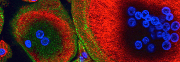
THE PRE-CLINICAL CHALLENGE IN 3D
INVITED SPEAKERS
INVITED SPEAKERS
 |
Alexandre Dufour
LECTURE TITLE: Signal processing challenges in quantitative 3D cell morphodynamics |
|
| Dr. Alexandre Dufour graduated in Artificial Intelligence & Pattern Recognition from University Pierre & Marie Curie (Paris, France) in 2004, and performed his doctoral research at Institut Pasteur Korea (Seoul, South Korea) from 2005 to 2007, where he co-founded the Image Mining group as a Junior Scientist. He then pursued his postdoctoral work in the Bioimage Analysis group at Institut Pasteur (Paris, France), and is now a permanent Staff Scientist in the group, leading the « cell deformation and motility » research team. Dr. Alexandre Dufour is a member of the Institute of Electrical and Electronics Engineers (IEEE), the IEEE Signal Processing Society, an associate member of the IEEE Bio-Imaging and Signal Processing Technical Committee, and also a board member of the Functional Live Microscopy development group (GdR 2588) of the French National Council for Scientific Research (CNRS). His teaching activities include the EMBO course « Imaging Parasite Infection » and the CNRS school on live microscopy «MiFoBio». | ||
|
Arman Rahman
LECTURE TITLE: Fast-Tracking Histopathology in Oncology via Image Analysis: Case Studies in Colorectal Cancer.
|
|
Dr Arman Rahman is a MD and obtained his PhD in the field of Immunology from Umea University, Sweden. His primary research area was mucosal immunology specially the adaptive and innateimmune involvement in inflammatory bowel diseases. Dr Rahman joined Dublin City University in 2008 as a senior post-doctoral fellow and worked extensively in the field of protein transport machineries and later on antibody engineering. Arman spent almost two years working with Dr John McCafferty in Cambridge University, UK on a collaborative project developing antibodies against therapeutic target. Arman has extensive experience in tissue-based detection techniques such as Immunohistochemistry, in situ hybridization etc. |
||
 |
Elia Formisano
LECTURE TITLE: Computational neuroimaging of human (auditory) perception and cognition |
|
| Full professor at the Department of Cognitive Neuroscience, Maastricht University; Scientific Director Maastricht Brain Imaging Center (MBIC); Member of the Maastricht Center for Systems Biology (MaCSBio). Elia Formisano received his MSc degree in Electronic Engineering in 1996 from the University of Naples (Italy) and his PhD from the national (Italian) program in Bioengineering in 2000. Thanks to an outgoing grant, in 1998-1999, he was a visiting research fellow at the Max Planck Institute for Brain Research in Frankfurt/Main (with Dr. Rainer Goebel). In January 2000, he was appointed Assistant Professor at Maastricht University (Faculty of Psychology and Neuroscience) where he is now Full Professor of Neural Signal Analysis. In 2008-2013, he has been Head of the Department of Cognitive Neuroscience. He is scientific director of the Maastricht Brain Imaging Center (MBIC) and Principal Investigator of the "Auditory Perception and Cognition" research group. His research aims at discovering the neural basis of human auditory perception and cognition by combining multimodal functional neuroimaging with methods of machine learning and computational modeling. He pioneered the use of ultra-high magnetic field (7 Tesla) functional MRI in neuroscience studies of audition. Methods development focuses on algorithms for unsupervised (e.g. independent component analysis) and supervised (e.g. multivariate classification and regression) learning. On these topics, he has published in high ranked journals, including Science, Neuron, PNAS, Current Biology and received prestigious funding, such as NWO VIDI (2005-2010) and NWO VICI (2013-2018). |
||
 |
Huw Summers
LECTURE TITLE: Cell demographics beyond the Normal |
|
| I obtained my BSc and PhD degrees in Physics at Cardiff University, U.K. and spent my early career on research in Optoelectronics focussed towards data storage and transmission. In 2007 I took the decision to take a major new direction and embarked on work in analytical techniques and computational modelling for cell population biology. My major interest is in understanding cell heterogeneity and my approach is a top-down systems one in which cell population dynamics are described using a language of mathematical probability and molecular level mechanisms are inferred from population wide distributions of individual cell characteristics rather than directly measured. This is the black box approach taken by Engineers in system control, where a mathematical transfer function describes the rules that govern the evolution of the system between states (e.g. proliferation of a cell population) rather than the underlying mechanisms. The experimental platforms with which we employ these approaches are flow cytometry and automated microscopy and the areas of interest include nanoparticle-cell interactions, immune system activation and cancer biology. | ||
 |
Julien Colombelli
LECTURE TITLE: Imaging model diseases in transparent organs |
|
| J.C. earned his Physics degree at the Universities of Paris XI (France) and Edinburgh (Scotland), and a MSc in Optical Engineering at the École Centrale Marseille (France) and the Politecnico di Milano (Italy). After a short period (2001-2002) developing optical instrumentation for Fiber Communications at Corecom in Milan (Italy), he moved to the Light Microscopy Group at the European Molecular Biology Laboratory (EMBL) in Heidelberg (2002-2008), to develop laser-based microscopy instruments. One of them, a Laser Nanosurgery platform, became a central tool in the projects of many groups in Europe focusing on Cell Biology, Biomechanics and Developmental Biology, and helped contributing to the understanding of cytoskeleton dynamics, mechanosensitive proteins or multicellular tissue mechanics. In 2008, J.C. took in charge the construction and management of the Advanced Digital Microscopy (ADM) Facility of the Institute for Research in Biomedicine (IRB Barcelona). The platform offers a wide range of state-of-the-art instruments to over 200 users in the area of Barcelona and is open to external scientists. ADM participates in the development of national (REMOA) and European (e.g. EuroBioImaging, Global BioImaging) research infrastructure and training networks and also pushes the establishment of a European network of BioImage Analysts (EuBIAS). The platform also develops research instruments and techniques, including Custom Lightsheet Microscopy for living organisms and transparent organs, or feedback Analysis-to-Acquisition workflows (Intelligent Imaging) on confocal, epifluorescence or Lightsheet systems, all dedicated to the design of custom solutions beyond the state-of-the-art to answer challenging questions related to Biomedicine, e.g. tumor growth, angiogenesis, Alzheimer disease, metastases proliferation, etc | ||
 |
Martin Meier
LECTURE TITLE: Bioimaging as a tool; from preclinical animal Models to translational Research |
|
| Dr. Meier started as a biologist with a degree from Ruhr-University Bochum, graduated in neuroscience at University of Bremen and specialized in MRI at that time. During his postdoc time his stay as an invited researcher at RIKEN in Japan was the greatest experience. With interim stops in bioindustry and the Leibniz Institute for Neurobiology in Magdeburg Dr. Meier became head of the Preclinical Imaging Center at the central animal Facility of Hannover Medical School, supporting biomedical research with the major in vivo imaging tools. His research is concerned with the application of imaging technology to research problems in animal models of human diseases. He is tightly connected to and involved in research in the field of cardiology, angiology, neurology, implantology and organ transplantation. He is a workgroup leader in the Cluster of Excellence REBIRTH and a principal investigator in the Virtual Institute MetBioMat: in vivo studies of biodegradable magnesium based implant materials. Over the years his approach has been to combine structural and functional imaging techniques with quantitative and molecular biomarkers to accurately characterize the tissue parameters involved. Using multimodal approaches, his group is carrying out studies in small animal models for diverse applications. He is also acting as the Chairman of the local chapter of the VBio, German Life Sciences Association, in Bremen and Lower Saxony |
||
 |
Ricardo Henriques
LECTURE TITLE: Democratizing live-cell high-speed super-resolution microscopy - Yes you can!
|
|
| I was born in Lisbon Portugal 1980, the youngest son of a Mathematician and a Nurse. Brother to a Plasma Physicist from the Lisbon Institute of Technology (IST) and to a Singing Professor at the Superior Music Conservatory of Aragón. Supporting no (good) skills for music when compared to my brother, I chose to pursue studies in Particle Physics at the Lisbon Faculty of Sciences (FCUL). After graduating, I became increasingly interested in the application of Optical Physics and Image Analysis into the field of Cell Biology. Since then I have been involved in research in and with Optical Microscopy. In 2008, I started a Ph.D. in the field of Super-Resolution Microscopy with the group of Dr. Musa Mhlanga, divided between South Africa (CSIR) and Portugal (IMM); during this time I became a long standing visiting scientist at Institute Pasteur Paris, having done most of my research there at the group of Dr. Christophe Zimmer. One of the major focuses of my Ph.D. was to democratize Super-Resolution Microscopy, providing analytical and optical tools allowing easy access to this new methodology. One of my publications QuickPALM presented an algorithm that easily enabled Super-Resolution analysis, rapidly becoming a standard in the field. During this time I consulted for companies such as Andor Technology, Olympus Microsopy and BitPlane, who have integrated part of the technology I have developed in their systems. In 2011, I have done a short postdoc at Institute Pasteur, studying the formation of the immunological synapse and its subversion by HIV-1 infection. In 2013 I became a research group leader at the University College London. As a group, our research is divided between cell biology, optical physics and biochemistry. We focus on biological problems that cannot be challenged with current imaging technology, and thus we set forth to develop the analytical and optical tools to answer these questions. Some of our favourite open questions surround figuring out how HIV-1 enters cells, probing and remodelling membranes. To do so, we are working on new classes of fluorescent probes and high-speed cell friendly Super-Resolution methods that although optimally designed to answer our focus questions, will have broad applications into Cell Biology research. | ||
 |
Ronit Satchi-Fainaro
LECTURE TITLE: Novel cancer models-based identification of targets for the rational design of precision nanomedicines | |
| Prof. Ronit Satchi-Fainaro (Ph.D.) is Head of the Cancer Angiogenesis & Nanomedicine Laboratory; Chair of the Department of Physiology & Pharmacology, Sackler School of Medicine. Prof. Satchi-Fainaro received her Bachelor of Pharmacy from the Hebrew University, Israel (1995) and her Ph.D. from the University of London, UK (1999). She then spent two years as postdoctoral fellow at Harvard University and Childrens Hospital Boston working with Judah Folkman on novel angiogenesis-targeted nanomedicines. In 2003, she was appointed Instructor in Surgery at Boston Childrens Hospital and Harvard Medical School and continues to have a Visiting Associate Professor position there to date. She joined Tel Aviv University in 2006.She is a leader in the field of nanomedicine and angiogenesis (cancer and vascular biology). She serves as advisor to several Israeli and International biotech and pharma companies, is President of the Israeli Society for Controlled Release, and is on the editorial boards of several biological and chemical journals. She has published more than 70 papers, 12 book chapters, edited 2 books, is named inventor on 30 patents, and was awarded numerous prestigious grants and prizes, among them Fulbright, Rothschild, Wingate, Alon, Young Investigator Award of the European Association for Cancer Research, 50 most influential women in Israel (Globes, Calcalist, TheMarker, Forbes), Juludan Prize for the Advancement of Technology in Medicine, the 2013 Teva Pharmaceutical Industries Founders Award for the Discovery of new molecular mechanisms and targets that would lead to new therapeutic approaches. Recently, she received the European Research Council (ERC) Consolidator Award and the Saban Family Foundation-Melanoma Research Alliance (MRA) Team Science Award.She has major expertise in tumor biology, tumor dormancy, angiogenesis, molecular imaging, non-invasive intravital imaging of animal models, personalized nanomedicines for cancer theranostics (therapy and diagnostics). Throughout, she has maintained an interest in understanding the biological rationale for the design of nanomedicines suitable for transfer into clinical testing. Her multi-disciplinary research laboratory focuses on basic research leading to the design of highly-selective targeting molecules integrating biology, chemistry, protein engineering, molecular imaging, computational approaches, material sciences and nanotechnology to selectively guide drugs into pathological sites. | ||
RUA DO CAMPO ALEGRE, 823 | 4150-180 PORTO - PORTUGAL | TEL +351 226 074 900 | EMAIL: BIOIMAGING2015@INEB.UP.PT
| ORGANIZED BY: | CO-ORGANIZED BY: | SUPPORT: | |||||
 |
 |
||||||