BIOIMAGING 2015
4TH INTERNATIONAL SYMPOSIUM IN APPLIED BIOIMAGING
THE PRE-CLINICAL CHALLENGE IN 3D
LAB SESSIONS
Each lab session has a limited number of seats. Registration based of first-come, first-served.
A. micro-CT image acquisition and analysis for biomedical research
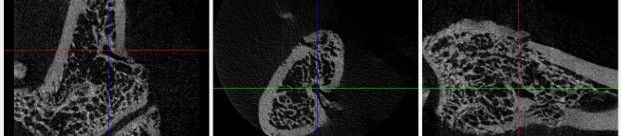
Micro-computed tomography (micro-CT) is a non-destructive technique that can be used to acquire three dimensional tissue images in small animals making it ideal for longitudinal preclinical studies. Although it has been classically applied for imaging mineralized skeletal tissues, with the use of contrast agents, soft tissue imaging is now possible.
In this lab session, the basic theoretical background of micro-CT image acquisition will be introduced and the students will be familiarized with micro-CT image acquisition and reconstruction in rodents. Using a rodent dataset, data will be analyzed and several parameters will be quantified such as tissue density, porosity and morphometric parameters using the specific software.
Instructors: Daniel Torre (Paralab)
B. Microscopy image processing and data extraction with ImageJ
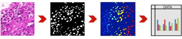
An image is worth a thousand... data points, and modern microscopy imaging techniques provides access to information rich images. Efficient data extraction from images requires however knowledge regarding different techniques, and the conditions of applicability and limitations of each one. Access to a basic "bag of tools" in image processing can be very useful to researchers interested in quantitative imaging.
This is a hands-on lab session, where the participants will learn and explore different image processing methodologies with relevance for microscopy. The well-established, and free, ImageJ software will be used to tackle case studies addressing common problems such as segmentation, morphometry and particle counting.
Instructors: Paulo Aguiar (INEB), António Pereira (IBMC)
C. Brain structure and function: a multimodal overview
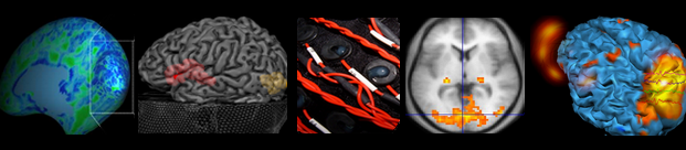
The technological advances in Brain Imaging systems and techniques revolutionized the modern Medicine. These are crucial for establishing the structure-function relationships in both healthy and pathological brain. The localization of specific areas of the brain that are affected by neurological or psychiatric disorders allows the development of new therapeutic strategies.
In this hands-on lab session, the participants will be able to learn and explore different data processing approaches. Well-established software will be used for data analysis of three main data types: functional MRI, structural MRI (e.g. Voxel Based Morphometry) and simultaneous EEG/fMRI.
Instructors: João Castelhano, João V. Duarte, Otília C. d'Almeida (IBILI)
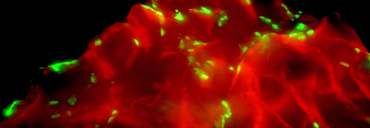
D. Perfusion cultures and Impedance measurements
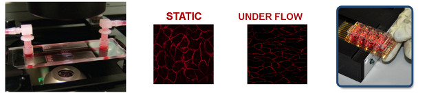
Lab-on-a-chip. Longterm-cultivation of endothelial cells under shear stress conditions in microfluidic-like channel chambers and monitoring changes in cell morphology with impedance measurements
Several adherent cell types are exposed to shear stress conditions in vivo, e.g. endothelial cells in a blood vessel. Culturing cells in vitro under perfusion conditions simulates this mechanical stimulus and induces a more physiological behavior. It has been previously shown that shear stress changes the cell morphology as well as crucial biological functions such as the barrier function of the cell.
Microfluidic-like chambers with defined channel geometries in combination with a perfusion system allow the long-term cultivation of cells under defined shear stress conditions. This provides an ideal in vitro setup to study endothelial and some epithelial cells in an in vivo-like environment. While shear stress induced morphological changes can be often easily detected under the microscope, changes for example in the barrier of the cells, are often harder to detect. Electric Cell Substrate impedance Sensing (ECIS) is a very sensitive, label-free method that allows to monitor and measure, even small changes in cell morphology, in real-time.
In this lab session the students will be introduced to:
- the use of a perfusion device in combination with microfluidic-like channel chambers that allow the long-term cultivation of cells under shear stress conditions. Changes in cell morphology can be subsequently observed under the microscope.
- real-time monitoring of cell growth/proliferation and barrier formation using an impedance measurement technique (ECIS).
Instructors: Tina Freisinger, Helga Wagner - IBIDI
RUA DO CAMPO ALEGRE, 823 | 4150-180 PORTO - PORTUGAL | TEL +351 226 074 900 | EMAIL: BIOIMAGING2015@INEB.UP.PT
| ORGANIZED BY: | CO-ORGANIZED BY: | SUPPORT: | |||||
 |
 |
||||||